
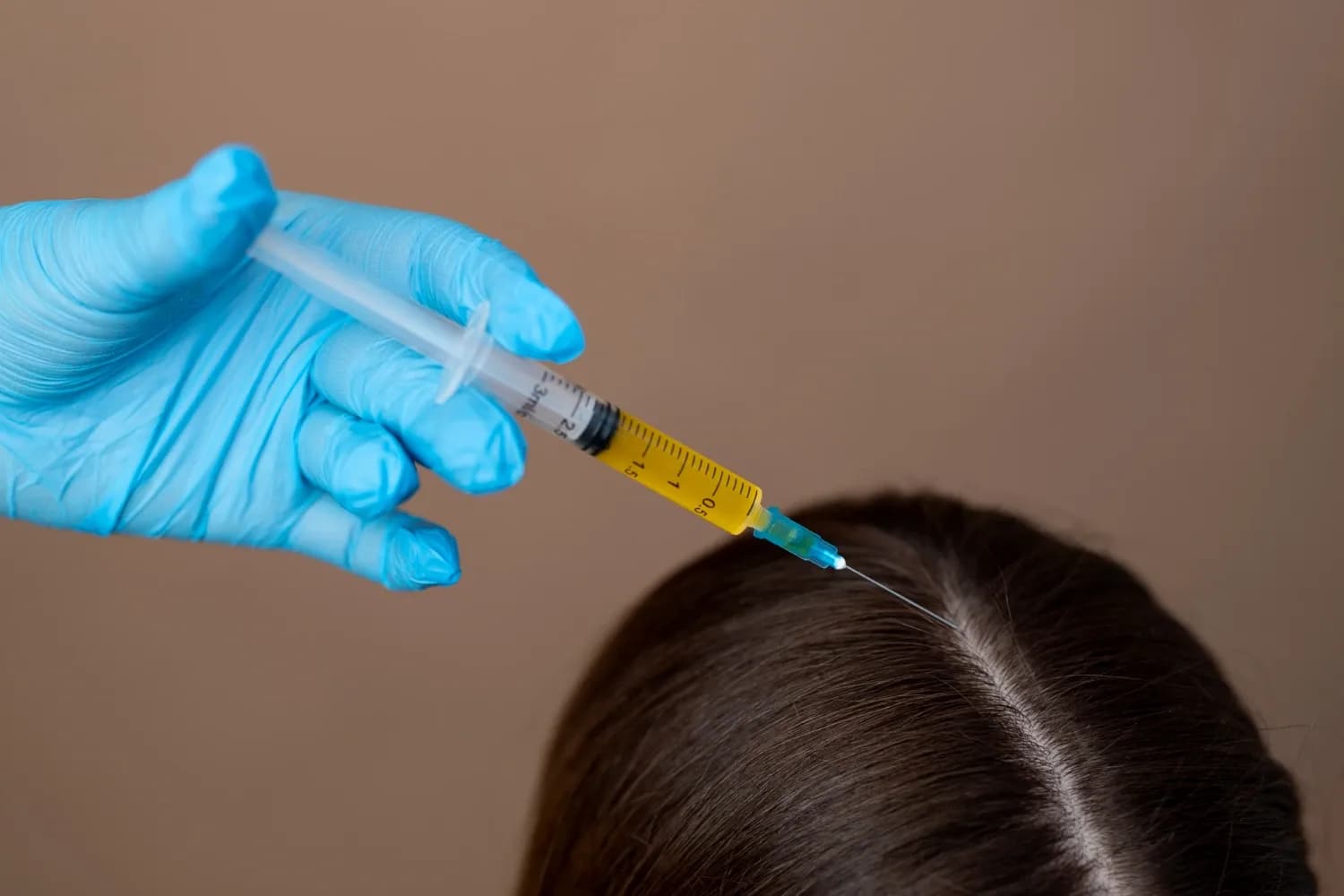
PRF vs Exosome: Which Regenerative Treatment Is Better?
Regenerative medicine now offers new hope for people struggling with hair loss and skin aging. Two treatments have gained significant attention in recent years.
Regenerative medicine now offers new hope for people struggling with hair loss and skin aging. Two treatments have gained significant attention in recent years. These are Platelet-Rich Fibrin (PRF) and exosome therapy. Both approaches use biological signals to stimulate repair and renewal. However, they work through very different biological mechanisms. This article compares PRF vs exosome therapy in detail. We examine their sources, compositions, mechanisms, clinical evidence, safety profiles, and practical applications. We also explain why promising laboratory findings do not automatically translate into proven clinical superiority.
What Is PRF?
PRF stands for Platelet-Rich Fibrin. It is an autologous blood preparation that contains platelets, fibrin, and growth factors. Doctors prepare it from the patient's own blood without anticoagulants.
PRF represents an advanced form of platelet-based regenerative therapy. Researchers developed it as an improvement over earlier platelet preparations. The key innovation involves eliminating anticoagulants during processing. This allows natural fibrin clot formation. The resulting material acts as both a biological scaffold and a sustained-release delivery system for healing signals.
How Is PRF Prepared?
Doctors draw blood from the patient. They spin it in a centrifuge without anticoagulants. The blood separates into layers. The fibrin matrix forms naturally. Clinicians then collect the PRF preparation for use.
The preparation process matters greatly for the final product. A clinician draws a small volume of blood from the patient's arm. The blood goes into tubes without any chemical anticoagulants. The tubes then undergo centrifugation at specific speeds and times. The exact parameters vary by protocol. Some protocols use lower speeds. Others use higher speeds. These differences affect platelet concentration and leukocyte content. After centrifugation, the blood separates into distinct layers. The middle layer contains the fibrin clot with trapped platelets and cells. Clinicians extract this layer for injection or topical application. Choukroun et al. (2001) first described this preparation method. They emphasized that the absence of anticoagulants allows natural clotting cascade activation. Dohan Ehrenfest et al. (2009) later classified different platelet concentrate types. They highlighted how preparation variables influence biological outcomes.
What Does PRF Contain?
PRF contains platelets, a fibrin matrix, growth factors, cytokines, and leukocytes. The exact composition depends on the preparation protocol.
PRF contains several biologically active components. Platelets form the core element. These cell fragments release growth factors upon activation. The fibrin matrix provides structural support. It also controls the release kinetics of signaling molecules. Growth factors include Platelet-Derived Growth Factor (PDGF), Vascular Endothelial Growth Factor (VEGF), and Transforming Growth Factor-beta (TGF-β). Cytokines add additional regulatory signals. Leukocytes may also be present depending on the centrifugation protocol. Some protocols intentionally include white blood cells. Others aim to minimize them. This variability means different PRF preparations may have different biological profiles.
How Does PRF Work?
PRF works through platelet activation, gradual growth factor release, and fibrin scaffold support. These processes stimulate tissue repair and cellular migration.
When clinicians inject PRF into tissue, the platelets encounter collagen and tissue factors. This contact triggers platelet activation. The platelets then release their granule contents. Growth factors and cytokines enter the surrounding tissue. The fibrin matrix controls this release. It prevents rapid dispersion of the signals. Instead, the tissue receives a sustained supply of regenerative molecules over several days. The fibrin also acts as a scaffold. Cells migrate into this matrix. New blood vessels begin to form. The combined effect supports tissue repair and renewal. In hair applications, these signals may influence hair follicle cells. Gentile and Garcovich (2020) reviewed PRF applications in alopecia. They noted that injectable PRF shows potential for improving hair density. However, they also pointed out that high-quality clinical studies remain limited. A systematic review by Gupta et al. (2021) identified only seven eligible dermatological studies on PRF. Three of these reported improvements in hair density and growth. The remaining studies showed less clear outcomes.
What Are Exosomes?
Exosomes are tiny extracellular vesicles that carry signaling molecules between cells. They enable intercellular communication and tissue regulation.
Exosomes represent a newer frontier in regenerative medicine. These nanoscale particles serve as biological messengers. They travel between cells and deliver functional cargo. Unlike cell-based therapies, exosomes contain no living cells. This makes them a cell-free regenerative approach.
What Are Exosomes Exactly?
Exosomes are extracellular vesicles measuring 30 to 150 nanometers. They carry microRNAs, proteins, lipids, and other signaling molecules. They differ from stem cells because they contain no nuclei or replication capacity.
Scientists classify exosomes as a subtype of extracellular vesicles. Cells release them through a specific biological process involving multivesicular bodies. The exosome membrane contains lipids and proteins from the parent cell. Inside, they carry a rich cargo of biologically active molecules. MicroRNAs form a key component. These small RNA molecules regulate gene expression in recipient cells. Proteins include enzymes, signaling molecules, and structural components. Lipids help stabilize the vesicle and facilitate cell entry. Pegtel and Gould (2019) described exosomes as critical mediators of intercellular communication. Tkach and Théry (2016) explained how cells use exosomes to exchange information across tissues. This communication system plays roles in development, immunity, and tissue repair.
Where Do Exosomes Come From?
Exosomes come from mesenchymal stem cells, adipose tissue, umbilical cord tissue, placental tissue, and dermal papilla cells. The source determines the exosome content.
Different cell types produce exosomes with different cargo profiles. Mesenchymal stem cell-derived exosomes have received the most research attention. These cells come from bone marrow, adipose tissue, or umbilical cord. Adipose-derived exosomes carry signals related to fat tissue biology. Umbilical-cord-derived exosomes come from perinatal tissue. Some products use placental sources. Dermal papilla cell exosomes specifically relate to hair follicle biology. Researchers have also explored exosomes from other cell types. The biological source matters because it determines which signaling molecules the exosomes contain. Stevens and Khetani (2023) emphasized that exosome heterogeneity poses a major challenge for clinical translation. Products from different sources may produce very different biological effects.
How Might Exosomes Support Hair Growth?
Exosomes may support hair growth through cell signaling, Wnt pathway modulation, VEGF-related angiogenesis, and dermal papilla cell stimulation. However, human clinical evidence remains early and heterogeneous.
Exosomes influence hair biology through multiple pathways. They deliver signals directly to hair follicle cells. The Wnt/β-catenin pathway plays a central role in hair development. Exosomes may modulate this pathway. They also carry VEGF-related signals. These promote blood vessel formation around follicles. Dermal papilla cells control hair growth cycling. Exosomes may influence these cells directly. Some research suggests anti-inflammatory effects. Inflammation contributes to certain types of hair loss. Exosomes may help reduce this. Hu et al. (2022) demonstrated that mesenchymal stem cell exosomes promote hair follicle development in preclinical models. Zhou et al. (2023) reviewed clinical studies and found promising improvements in hair density and thickness. However, they noted substantial variation in exosome sources, doses, routes, and outcome measures across studies.
PRF vs Exosome: What Is the Difference?
PRF comes from the patient's own blood and contains platelets in a fibrin matrix. Exosomes come from processed biological sources and contain nanoscale signaling vesicles. They differ in source, composition, mechanism, and standardization.
The fundamental difference between PRF and exosomes lies in their biological nature. PRF is a whole biological preparation from autologous blood. Exosomes are isolated extracellular vesicles from various sources. This distinction affects every aspect of their clinical use.
PRF vs Exosome at a Glance
PRF uses autologous blood with a fibrin scaffold and gradual growth factor release. Exosomes use donor-derived vesicles with direct intercellular signaling. Both lack strong standardized clinical evidence for hair loss.
Feature | PRF | Exosomes |
Biological source | Patient's own blood | Usually processed biological source |
Main components | Platelets, fibrin, growth factors, cells | Extracellular vesicles, signaling cargo |
Autologous | Yes | Usually no |
Fibrin matrix | Yes | No |
Signaling mechanism | Growth-factor release and tissue repair | Intercellular molecular signaling |
Release profile | Relatively sustained through fibrin | Depends on product and formulation |
Standardization | Varies by preparation protocol | Varies substantially by source and manufacturing |
Hair-loss evidence | Emerging | Emerging |
Main scientific limitation | Protocol heterogeneity and limited trials | Product heterogeneity and limited high-quality clinical data |
What Differs in Source and Composition?
PRF uses autologous whole blood. Exosomes use isolated vesicles from donor or cultured cells. This difference affects consistency, safety, and biological predictability.
PRF derives entirely from the patient's own blood. This eliminates concerns about donor compatibility or disease transmission. However, it also means each preparation reflects the individual patient's biology. Platelet counts vary between people. Growth factor levels differ. Age, health status, and medications all influence PRF quality. Exosomes come from external biological sources. Manufacturers process these sources to isolate vesicles. The starting material may be stem cell cultures, tissue extracts, or other biological preparations. This allows potential for standardization. However, it also introduces variability between products. Different manufacturers use different isolation methods. Different cell sources produce different exosome cargo. Bansal and Hughes (2021) documented how PRF preparation protocols significantly affect platelet concentration and growth factor levels. Stevens and Khetani (2023) similarly showed that exosome products vary enormously in vesicle concentration, purity, and biological activity.
How Does Growth-Factor Release Differ?
PRF releases growth factors gradually through its fibrin matrix. Exosomes deliver pre-packaged signaling molecules through vesicle fusion with cells. These are fundamentally different delivery mechanisms.
PRF functions as a biological delivery system. The fibrin matrix traps growth factors and platelets. Over time, the matrix degrades. This releases the active components gradually. The tissue receives a sustained signal over days or weeks. Exosomes work differently. They do not primarily release free growth factors. Instead, they fuse with target cells or are taken up by them. They deliver their cargo directly into the recipient cell. This represents active intercellular communication rather than passive factor release. The distinction matters clinically. PRF provides a prolonged but diffuse signal. Exosomes provide targeted but potentially shorter-lived signals. Scientists should not treat these mechanisms as interchangeable.
What About Biological Signaling?
PRF triggers platelet-mediated regenerative cascades. Exosomes enable vesicle-mediated cellular communication. Both may activate similar downstream repair pathways but through different starting mechanisms.
PRF initiates the classical wound healing cascade. Platelets release PDGF, VEGF, TGF-β, and other factors. These recruit immune cells, fibroblasts, and endothelial cells. The result is a coordinated repair response. Exosomes bypass some of these early steps. They deliver signals that may directly influence cell behavior. MicroRNAs can alter gene expression. Proteins can activate specific pathways. Both approaches may ultimately stimulate collagen production, angiogenesis, and cellular renewal. However, they reach these endpoints through different biological routes. This explains why some patients respond better to one treatment than the other.
Why Does Standardization Matter?
PRF standardization depends on centrifugation speed, time, and tube type. Exosome standardization depends on source, isolation method, and vesicle characterization. Without standardization, comparing treatments becomes scientifically unreliable.
Standardization represents a critical challenge for both therapies. PRF protocols vary in centrifugation parameters. Speed, time, and relative centrifugal force all affect the product. Tube design influences clot formation. Some protocols include leukocytes. Others exclude them. These variations produce biologically different preparations. Exosome products face even greater standardization challenges. Manufacturers use different isolation techniques. Ultracentrifugation, precipitation, and chromatography yield different results. Vesicle concentration varies. Purity varies. Cargo characterization remains incomplete. Scientists cannot reliably compare "PRF" with "exosomes" without specifying exact protocols and products. A specific PRF protocol may outperform a specific exosome product, or vice versa. General comparisons lack scientific validity.
PRF vs Exosome for Hair Loss: Which Shows More Promise?
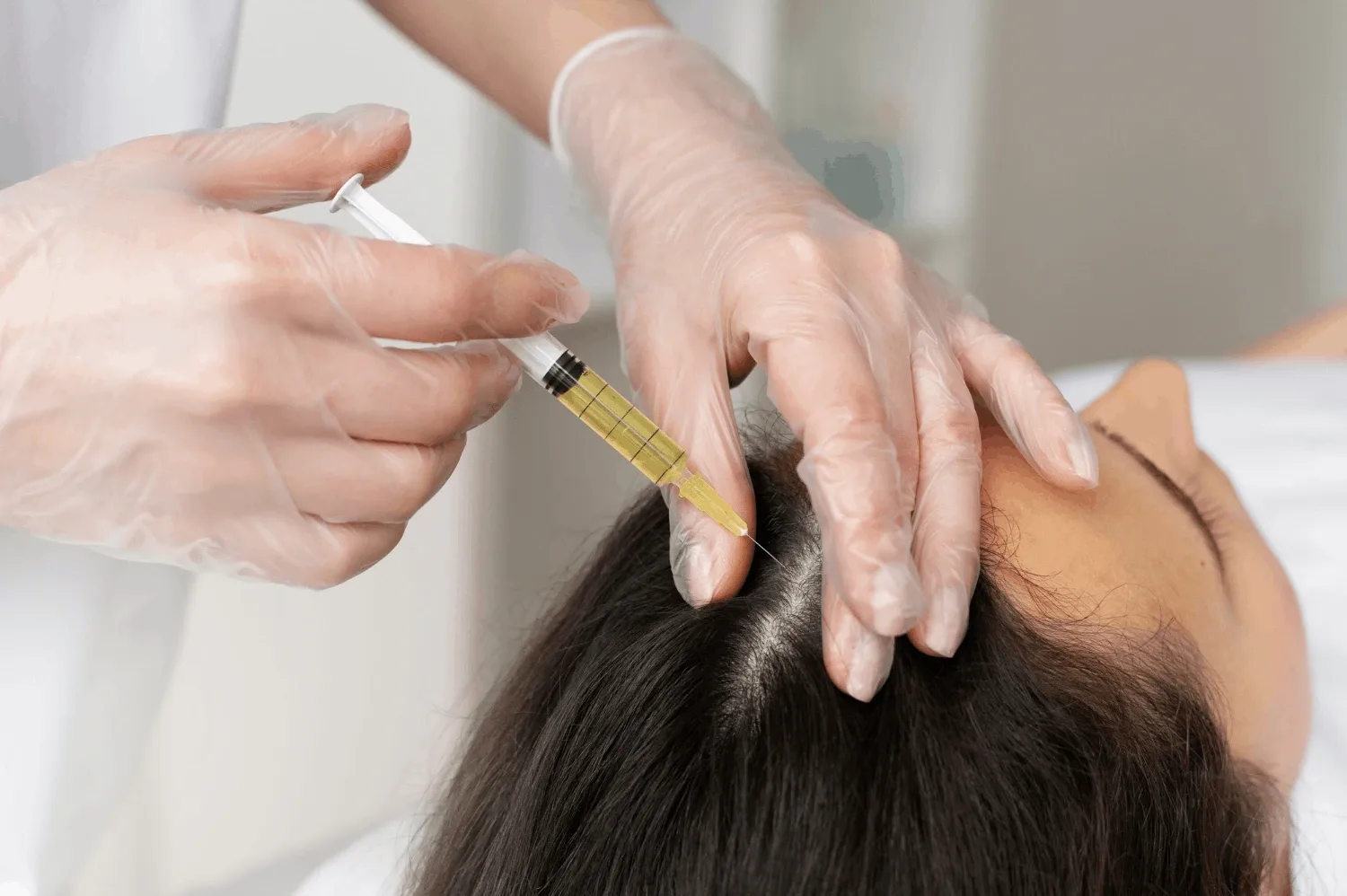
Both PRF and exosomes show emerging evidence for hair loss. Neither has established definitive superiority. PRF has a longer history in platelet medicine. Exosomes have more recent but heterogeneous clinical data.
Hair loss affects millions of people worldwide. Androgenetic alopecia remains the most common cause. Both PRF and exosomes have entered clinical practice as potential treatments. However, the evidence base for each remains in development.
How Does PRF Perform for Androgenetic Alopecia?
PRF may improve hair density and thickness in some patients with androgenetic alopecia. However, high-quality controlled trials remain limited.
Androgenetic alopecia involves progressive hair follicle miniaturization. Dihydrotestosterone shortens the anagen growth phase. Follicles produce thinner, shorter hairs over time. PRF aims to counteract this process. Growth factors in PRF may stimulate follicle stem cells. VEGF may improve blood supply to the follicle. PDGF may support dermal papilla cell function. Gentile and Garcovich (2020) reported that PRF injections increased hair density in treated areas. However, they noted significant variability between patients. Gupta et al. (2021) conducted a systematic review of PRF in dermatology. They found only seven eligible studies. Three showed improvements in hair density and growth. Four showed less definitive results. All studies had methodological limitations. Sample sizes were small. Control groups were often absent. Follow-up periods were short. Gkini et al. (2014) studied PRP, a related therapy, and found measurable increases in hair density. PRF builds on this foundation but lacks equivalent trial volume.
How Do Exosomes Perform for Androgenetic Alopecia?
Early clinical studies suggest exosomes may increase hair density, thickness, and scalp coverage. However, study quality varies, and long-term data remain scarce.
Exosomes target hair loss through multiple signaling pathways. They may influence dermal papilla cells directly. They may modulate Wnt/β-catenin signaling. This pathway regulates hair follicle development and cycling. They may enhance VEGF expression. This promotes angiogenesis around follicles. They may also exert anti-inflammatory effects. Zhou et al. (2023) identified 11 clinical studies of exosome-based hair treatments. They reported promising improvements in hair density, thickness, and scalp coverage. However, they highlighted substantial variation in exosome sources, doses, administration routes, and outcome measures. Some studies used adipose-derived exosomes. Others used umbilical-cord sources. Dosing ranged widely. Delivery methods included injection, microneedling, and topical application. This heterogeneity makes it impossible to draw firm conclusions about efficacy. O'Connell et al. (2021) reviewed exosome therapy in aesthetic medicine. They described the approach as promising but emphasized the need for stronger clinical validation.
Which Works Better for Hair Loss?
Current science cannot definitively declare either treatment superior for hair loss. Both have biological plausibility. Both have emerging clinical evidence. Patient-specific factors and product quality likely matter more than the treatment category.
Comparing PRF and exosomes for hair loss requires careful nuance. Evidence maturity slightly favors PRF in the broader context of platelet-based therapies. PRP, the predecessor to PRF, has more published studies. However, PRF-specific research remains limited. Exosome research is growing rapidly but remains early. Biological plausibility exists for both. PRF provides a sustained release of multiple growth factors. Exosomes provide targeted cellular signaling. Patient-specific suitability varies. Some patients may respond better to autologous preparations. Others may benefit more from concentrated exosome products. Product consistency affects outcomes more than treatment category. A well-prepared PRF may outperform a poor exosome product. A high-quality exosome preparation may surpass a poorly executed PRF treatment. Safety considerations also differ. PRF carries minimal disease transmission risk. Exosome products require rigorous quality control. Clinicians should discuss established evidence-based therapies alongside these regenerative options. Minoxidil, finasteride, and low-level laser therapy have stronger clinical evidence. These should remain part of comprehensive hair loss management.
PRF vs Exosome for Skin Rejuvenation: Which Is More Effective?
Both PRF and exosomes show potential for skin rejuvenation. PRF emphasizes collagen stimulation through sustained growth factor release. Exosomes emphasize cellular communication and repair signaling. The better choice depends on the specific skin concern and product quality.
Skin aging involves collagen loss, elastin degradation, and reduced cellular turnover. Both PRF and exosomes aim to reverse these changes. However, their mechanisms suit different rejuvenation goals.
How Does PRF Help Skin?
PRF stimulates collagen production, supports tissue remodeling, and improves skin thickness and elasticity. Clinicians use it around the eyes, for scars, and for general facial rejuvenation.
PRF promotes skin renewal through the same mechanisms that drive wound healing. Growth factors stimulate fibroblasts. These cells produce new collagen and elastin. The fibrin scaffold supports tissue organization. Over time, skin thickness increases. Elasticity improves. Fine lines may soften. Clinicians apply PRF in several ways. They inject it into specific areas. They combine it with microneedling. They apply it topically after laser treatments. The periocular region responds well to PRF. Thin under-eye skin benefits from collagen stimulation. Acne scars may improve through tissue remodeling. Suga et al. (2015) demonstrated that fibrin matrices support organized tissue repair. This explains why PRF produces gradual, natural-looking improvements rather than dramatic immediate changes.
How Do Exosomes Help Skin?
Exosomes may enhance cellular communication, reduce inflammation, and promote skin repair. Clinicians often combine them with microneedling for enhanced delivery.
Exosomes influence skin biology through intercellular signaling. They may stimulate fibroblast activity. They may modulate inflammatory responses. Chronic inflammation accelerates skin aging. Exosomes may help regulate this process. They may also support barrier repair. The lipid content of exosomes may enhance skin hydration. Microneedling creates controlled microchannels in the skin. Applying exosomes after microneedling may improve penetration. The combination aims to maximize delivery to deeper skin layers. However, the optimal exosome concentration, source, and application protocol remain under investigation. Marotta et al. (2022) reviewed regenerative approaches in dermatology. They noted that exosome skin applications show early promise. However, they called for more rigorous clinical trials with standardized products.
Which Is Better for Skin Rejuvenation?
Neither treatment is universally better for skin rejuvenation. PRF suits patients wanting autologous collagen stimulation. Exosomes suit patients wanting concentrated signaling molecules. The choice depends on skin concern, desired outcome, delivery method, and product quality.
Selecting between PRF and exosomes for skin requires individualized assessment. PRF offers the advantage of autologous sourcing. Patients who prefer treatments from their own biology may favor this option. The fibrin matrix provides sustained activity. Results develop gradually over months. Exosomes offer concentrated signaling potential. A single treatment may deliver high levels of active molecules. However, the effects may be shorter-lived. The skin concern matters. PRF may work better for volume loss and deep tissue support. Exosomes may work better for surface texture and inflammation. Delivery method affects outcomes. Injection places material deeper. Microneedling distributes it more superficially. Product quality determines biological activity. A poorly prepared PRF or low-quality exosome product will not produce good results regardless of the treatment category.
PRF vs Exosome: How Is Each Treatment Performed?
PRF involves blood draw, centrifugation, and injection or topical application. Exosome treatment involves product selection, skin preparation, and delivery via injection or microneedling. Both require medical oversight.
Understanding the treatment process helps patients set realistic expectations. Both procedures are minimally invasive. However, they differ in preparation and delivery.
How Is PRF Treatment Performed?
The clinician assesses the patient, draws blood, spins it in a centrifuge, prepares the PRF, and injects or applies it. Post-treatment monitoring follows.
The PRF treatment process begins with patient assessment. The clinician reviews medical history. They examine the treatment area. They discuss goals and expectations. The actual procedure starts with blood collection. A clinician draws 10 to 20 milliliters of blood from a arm vein. The blood goes into special tubes. These tubes contain no anticoagulants. The tubes then enter a centrifuge. Spinning separates blood components by density. Red blood cells sink to the bottom. The fibrin clot forms in the middle layer. This layer contains the PRF. The clinician extracts this material carefully. They may inject it directly into the scalp or skin. Alternatively, they may apply it topically after microneedling. The entire process takes 30 to 60 minutes. Patients may experience mild redness or swelling. These effects typically resolve within 24 to 48 hours.
How Is Exosome Treatment Performed?
The clinician assesses the patient, selects an appropriate exosome product, prepares the skin or scalp, and delivers the exosomes through injection or microneedling. Post-treatment care instructions follow.
Exosome treatment also begins with assessment. The clinician evaluates the patient's condition. They select an exosome product based on the treatment goal. Product selection requires knowledge of the specific formulation. Not all exosome products are equivalent. The clinician prepares the treatment area. They may apply topical numbing cream. They clean the skin or scalp thoroughly. Delivery occurs through injection or microneedling. Some protocols use both. Injection places exosomes deeper into tissue. Microneedling distributes them across a broader surface area. The clinician may combine exosomes with other treatments. Post-treatment care includes avoiding sun exposure and harsh products for several days. Patients should follow all aftercare instructions carefully.
Why Does Delivery Method Matter?
Injection places material deep into tissue. Microneedling creates surface channels for absorption. Topical application alone has limited penetration. The delivery method affects treatment depth and outcomes.
The way clinicians deliver regenerative products significantly affects results. Injection ensures placement at the target depth. For hair loss, this means near the hair follicle bulbs. For skin, this means the dermis. However, injection covers limited area. Microneedling treats larger surface areas. The microchannels allow product entry. However, penetration depth is shallower. Combining both methods may optimize coverage and depth. Topical application without microneedling rarely achieves adequate penetration. The stratum corneum blocks most molecules. Exosomes and PRF components cannot easily cross this barrier alone. Protocol-dependent outcomes mean that the same product may produce different results with different delivery methods.
PRF vs Exosome: What Are the Benefits and Limitations?
PRF offers autologous sourcing and a fibrin scaffold but has biological variability. Exosomes offer concentrated signaling but lack standardization and long-term data. Both have strengths and weaknesses.
Every medical treatment involves trade-offs. PRF and exosomes each offer distinct advantages. They also face specific limitations.
What Are the Potential Benefits of PRF?
PRF uses the patient's own blood, requires no external donor, provides a fibrin scaffold, releases growth factors gradually, and involves relatively straightforward preparation.
PRF offers several practical and biological advantages. The autologous source eliminates compatibility concerns. The body recognizes its own material. This reduces allergic reaction risk. No external biological donor is needed. The fibrin scaffold provides structural support. It also controls release kinetics. Growth factors emerge gradually rather than all at once. This mimics natural healing patterns. Preparation requires only a centrifuge and standard blood collection supplies. Most clinics can perform the procedure without specialized equipment. The active components come from the patient's own biology. This appeals to patients who prefer natural treatment approaches.
What Are the Limitations of PRF?
PRF requires blood collection, shows patient-to-patient variability, lacks protocol uniformity, and has limited high-quality hair-loss research. Results vary between individuals.
PRF also has significant limitations. The procedure requires venipuncture. Some patients dislike needles or feel anxious about blood draws. Biological variability affects every preparation. A patient with low platelet counts produces less potent PRF. Age reduces growth factor levels. Chronic illness alters blood composition. Protocol differences between clinics create inconsistent products. One clinic's PRF may differ substantially from another's. High-quality PRF-specific research for hair loss remains sparse. Most studies are small and uncontrolled. Results vary widely between patients. Some see noticeable improvement. Others see minimal change. Predicting individual response remains difficult.
What Are the Potential Benefits of Exosomes?
Exosomes contain high concentrations of signaling molecules, offer a cell-free approach, show potentially broad biological activity, and may allow standardized formulations.
Exosomes offer unique advantages. They contain concentrated signaling molecules. A small volume may deliver high biological activity. The cell-free approach avoids concerns about cell viability or tumorigenicity. Exosomes cannot replicate or transform. They simply deliver signals and degrade. The biological activity may be broad. A single exosome preparation can influence multiple pathways. This includes angiogenesis, inflammation, and cellular renewal. Manufacturing processes may eventually standardize products. Unlike PRF, which varies by patient, exosome products could achieve batch-to-batch consistency. This would improve predictability and research quality.
What Are the Limitations of Exosomes?
Exosomes come from variable biological sources, use inconsistent manufacturing methods, lack universal dosing standards, and have limited long-term human data. Regulatory and quality concerns also exist.
Exosome therapy faces substantial challenges. Different biological sources produce different products. A mesenchymal stem cell exosome differs from an adipose-derived exosome. Manufacturing methods vary between companies. Some use ultracentrifugation. Others use precipitation or filtration. These methods yield different vesicle populations. No universal dosing standards exist. Clinicians do not know the optimal concentration, volume, or frequency. Long-term human safety data remain limited. Most studies follow patients for only a few months. Regulatory frameworks for exosome products are still developing. Some products may not meet rigorous quality standards. Product sourcing and manufacturing controls require careful scrutiny. Stevens and Khetani (2023) emphasized that exosome hair therapy needs stronger clinical validation and better standardization before becoming a routine treatment.
Are PRF and Exosome Treatments Safe?
Both treatments are generally safe when performed by qualified providers. PRF carries typical injection risks. Exosomes carry additional product-quality risks. Proper sourcing and sterile technique matter greatly.
Safety represents a primary concern for any medical procedure. Both PRF and exosomes have favorable safety profiles in general. However, specific risks differ between the two.
How Safe Is PRF?
PRF causes mild injection-related effects like redness, swelling, bruising, and temporary tenderness. Serious complications are rare. The autologous source minimizes immunogenicity risk.
PRF safety data come from extensive use in dentistry, orthopedics, and aesthetic medicine. Common side effects relate to the injection itself. Patients may experience redness at the injection site. Swelling typically peaks within 24 hours. Bruising occurs occasionally, especially in thin skin areas. Temporary tenderness lasts a few days. These effects are self-limiting. They resolve without intervention. The autologous source minimizes immunological risk. The body does not reject its own blood components. Allergic reactions are virtually absent. Infection risk is low when clinicians use sterile technique. Serious complications like vascular occlusion or nerve injury are rare. They require improper injection technique rather than product-related toxicity.
How Safe Are Exosomes?
Exosomes cause injection or microneedling effects, possible local inflammation, and infection risk if quality control is inadequate. Product sourcing and manufacturing quality are critical safety factors.
Exosome safety depends heavily on product quality. Injection-related effects mirror those of PRF. Redness, swelling, and tenderness occur commonly. Local inflammation may develop. This usually resolves quickly. Infection risk exists if products or procedures lack adequate control. Non-sterile products or improper handling can introduce pathogens. The biological source matters. Allogeneic or xenogeneic sources require rigorous screening. Manufacturing quality determines purity. Contaminants like endotoxins or residual cells may cause adverse reactions. Medical oversight ensures appropriate patient selection and technique. Patients should verify product documentation. They should ask about source, manufacturing, and sterility testing.
Why Does Product Quality Matter?
Sterility, source documentation, manufacturing controls, product characterization, storage conditions, and traceability all affect safety and efficacy. Poor quality products produce poor or dangerous outcomes.
Product quality determines both safety and results. Sterile preparation prevents infection. Source documentation ensures the biological origin is known and appropriate. Manufacturing controls maintain consistency. Product characterization verifies vesicle concentration and purity. Storage conditions preserve biological activity. Exosomes may degrade if stored improperly. Traceability allows investigation if problems arise. Patients should seek treatments from reputable providers. They should ask about product specifications. They should avoid products with unclear origins or inadequate documentation.
PRF vs Exosome: How Much Clinical Evidence Exists?
Both treatments have emerging evidence. PRF benefits from a longer history in platelet medicine but lacks large hair-loss trials. Exosomes have growing clinical data but remain early in development. Neither has definitive superiority evidence.
Evidence quality determines medical confidence. Both PRF and exosomes need more research. However, their current evidence bases differ in character.
What Evidence Supports PRF?
Systematic reviews, prospective studies, and small clinical cohorts support PRF. However, controlled trials are limited. Protocol standardization remains needed.
PRF research spans multiple medical fields. Dental and orthopedic applications have the longest history. Dermatological and hair applications are newer. Systematic reviews have begun to appear. Gupta et al. (2021) reviewed PRF in dermatology. They found encouraging but limited data. Prospective studies show trends toward improvement. Small clinical cohorts report positive outcomes. However, large randomized controlled trials are scarce. Most studies lack control groups. Blinding is difficult because PRF preparation is visible. Sample sizes are typically under 50 patients. Follow-up periods range from 3 to 12 months. Longer-term data are largely absent. Protocol differences between studies prevent meta-analysis. Standardized preparation and dosing would strengthen the evidence base considerably.
What Evidence Supports Exosomes?
Preclinical studies, early clinical trials, prospective studies, and systematic reviews support exosomes. However, long-term follow-up is limited.
Exosome research is accelerating. Preclinical studies in cell culture and animal models show biological activity. Early clinical trials in humans report promising results. Prospective studies track outcomes over time. Systematic reviews have begun to consolidate findings. Zhou et al. (2023) identified 11 clinical studies of exosome hair therapy. They found improvements in hair density, thickness, and coverage. However, they noted significant methodological concerns. Study designs varied. Exosome sources differed. Dosing was inconsistent. Outcome measures were not standardized. Long-term follow-up was generally short. Most studies tracked patients for 3 to 6 months. Durability of results beyond one year remains unknown. O'Connell et al. (2021) called for larger, longer, and better-controlled studies.
What Does Current Research Actually Show?
Current research shows biological plausibility for both treatments. It does not prove clinical superiority of either. Improvement in some studies does not equal proof of efficacy. Small samples and short follow-up limit conclusions.
Separating biological evidence from clinical evidence is essential. Laboratory studies show that PRF contains growth factors. They show that exosomes carry signaling molecules. These findings establish biological plausibility. However, plausibility does not guarantee clinical effectiveness. Human studies must demonstrate that these biological properties translate into measurable patient benefits. Current research shows improvement in some treated patients. However, improvement does not equal proof. Natural variation, placebo effects, and concurrent treatments may explain some findings. Sample sizes in existing studies are too small to detect modest effects reliably. Follow-up periods are too short to assess durability. Heterogeneous products make direct comparisons impossible. A study using one PRF protocol and one exosome product cannot generalize to all PRF or all exosomes.
What Should Future Studies Examine?
Future studies need randomized controlled trials, direct PRF-versus-exosome comparisons, standardized doses, standardized delivery methods, objective measurements, long-term safety data, and patient-reported outcomes.
The research agenda for both treatments is clear. Randomized controlled trials provide the strongest evidence. These should compare active treatment to placebo or established therapy. Direct PRF-versus-exosome comparisons would answer the superiority question. However, such studies must specify exact products and protocols. Standardized treatment doses would allow replication. Standardized delivery methods would reduce protocol variability. Objective hair-density measurements using standardized photography or trichoscopy would improve accuracy. Hair-shaft diameter measurements would capture caliber changes. Long-term safety monitoring would identify delayed adverse effects. Patient-reported outcomes would capture satisfaction and quality of life. Only through such rigorous research can clinicians make evidence-based recommendations.
PRF vs Exosome: Which Treatment Is Right for You?
The right treatment depends on your specific condition, goals, and preferences. PRF suits patients wanting autologous therapy. Exosomes suit patients interested in emerging concentrated signaling approaches. Neither should replace proper diagnosis.
Individualized medicine requires matching treatment to patient. Neither PRF nor exosomes is right for everyone. Proper patient selection improves outcomes and satisfaction.
When Should You Consider PRF?
Consider PRF for early or moderate hair thinning, if you want an autologous regenerative treatment, if you prefer platelet-derived growth factor therapy, or for selected skin rejuvenation applications.
PRF fits specific patient profiles well. Early or moderate hair thinning responds better than advanced baldness. The follicles are still present but miniaturized. Regenerative signals may reactivate them. Patients who prefer treatments from their own biology often choose PRF. This eliminates concerns about donor material. Patients interested in growth factor therapy specifically may favor PRF. The preparation contains multiple growth factors in a natural matrix. Selected skin rejuvenation applications also suit PRF. Under-eye hollows, fine lines, and acne scars may improve. Patients should have realistic expectations. PRF produces gradual improvement over months. It does not create dramatic overnight changes.
When Should You Consider Exosomes?
Consider exosomes if you are interested in emerging regenerative approaches, selected hair restoration protocols, microneedling-assisted treatments, and if you understand the current evidence limitations.
Exosomes appeal to patients interested in cutting-edge therapies. The science of extracellular vesicles is advancing rapidly. Patients who want concentrated signaling molecules may prefer exosomes. A small volume can deliver high biological activity. Microneedling-assisted delivery suits patients who want surface-wide treatment. Exosomes distribute well across treated areas. However, patients must understand the evidence limitations. Exosomes are promising but not proven. Results are not guaranteed. Products vary in quality. Patients should ask detailed questions about the specific product being used.
When Should Neither Treatment Be the First Step?
Neither treatment should come first for unexplained sudden hair loss, suspected nutritional or endocrine causes, active scalp disease, scarring alopecia without specialist assessment, or advanced follicle loss where regenerative treatment cannot recreate absent follicles.
Regenerative treatments are not appropriate for all conditions. Unexplained sudden hair loss requires diagnosis first. Telogen effluvium, alopecia areata, and other conditions need specific treatment. Nutritional deficiencies require supplementation. Endocrine disorders need hormonal management. Active scalp diseases like psoriasis or severe seborrheic dermatitis need control before any cosmetic procedure. Scarring alopecia destroys follicles permanently. Regenerative treatments cannot restore destroyed follicles. Advanced baldness with no remaining follicles cannot respond to biological signaling. There is nothing left to stimulate. In these cases, hair transplantation or other surgical options may be more appropriate.
Why Does Diagnosis Come Before Regenerative Treatment?
Proper diagnosis identifies the alopecia type, assesses scalp health, evaluates follicular miniaturization, considers established treatments, and enables individualized planning.
Diagnosis forms the foundation of effective hair loss management. Different alopecia types require different approaches. Androgenetic alopecia responds to antiandrogens and growth stimulators. Alopecia areata requires immunomodulatory therapy. Telogen effluvium resolves when the trigger disappears. Scalp health affects treatment success. Inflammation, infection, or poor circulation limit results. Evaluating follicular miniaturization determines whether regenerative treatment is viable. If follicles are merely miniaturized, stimulation may help. If follicles are gone, stimulation cannot help. Established treatments with strong evidence should be considered first or alongside regenerative options. An individualized treatment plan addresses the specific cause, severity, and patient goals. Regenerative therapy should fit within this broader plan, not replace it.
Can PRF and Exosomes Be Combined?
Some clinicians combine PRF and exosomes theoretically. PRF provides an autologous fibrin environment. Exosomes add concentrated signaling. However, direct comparative evidence for combination therapy is lacking. Combining treatments increases cost and complexity without proven additive benefit.
Combination therapy is common in medicine. Clinicians often use multiple treatments with complementary mechanisms. PRF and exosomes might theoretically complement each other.
What Is the Rationale for Combination Therapy?
PRF provides an autologous fibrin-based regenerative environment with sustained growth factor release. Exosomes provide additional extracellular-vesicle signaling. The mechanisms may complement each other.
The theoretical rationale for combining PRF and exosomes is biologically plausible. PRF creates a local regenerative environment. The fibrin matrix supports cell migration. Growth factors stimulate repair processes. This environment might enhance exosome uptake or activity. Exosomes could add signals that PRF lacks. They carry microRNAs and specific proteins. These might activate pathways that platelet factors do not reach. The combination might produce synergistic effects. However, synergy requires proof. Biological plausibility does not establish clinical benefit.
What Does Evidence Say About Combining Them?
No high-quality studies directly compare combination therapy to either treatment alone. Clinicians should not assume combination is automatically better. Controlled studies are needed.
Despite the theoretical rationale, evidence for combining PRF and exosomes is essentially absent. No randomized trials compare the combination to PRF alone or exosomes alone. Case reports and anecdotal observations cannot establish superiority. They may reflect placebo effects, natural variation, or concurrent treatments. Combining therapies increases cost. Patients pay for both products and procedures. Complexity increases. Preparation time extends. Recovery may involve more variables. Without proven additive benefit, combination therapy remains experimental. Patients should understand this before choosing combination approaches.
PRF vs Exosome vs PRP: How Do They Compare?
PRF, PRP, and exosomes are three distinct regenerative approaches. PRF and PRP are autologous platelet preparations that differ in anticoagulant use and fibrin content. Exosomes are cell-free vesicles from various sources. PRP has the most clinical history. PRF offers a fibrin scaffold. Exosomes offer concentrated signaling.
Patients and clinicians often compare three regenerative options. Understanding all three helps informed decision-making.
How Do PRF and PRP Differ?
PRF uses no anticoagulants and forms a natural fibrin matrix. PRP uses anticoagulants and remains liquid. PRF releases growth factors more gradually. PRP allows immediate release.
PRF evolved from PRP. The key difference is anticoagulant use. PRP preparation requires anticoagulants like citrate. These prevent clotting during processing. The final product remains liquid. Clinicians activate PRP before injection. This triggers immediate growth factor release. PRF avoids anticoagulants. The blood clots naturally during centrifugation. The fibrin matrix traps platelets. Growth factors release gradually as the matrix degrades. PRP provides a burst of signals. PRF provides sustained signals. Biological composition also differs. PRF contains leukocytes in many protocols. PRP may or may not contain them. The fibrin scaffold in PRF adds structural support that PRP lacks.
How Do PRP and Exosomes Differ?
PRP is an autologous platelet product with immediate growth factor release. Exosomes are cell-free extracellular vesicles with intercellular signaling. They differ in source, mechanism, and clinical evidence.
PRP and exosomes differ fundamentally. PRP comes from the patient's blood. Exosomes come from external sources. PRP contains living platelets. Exosomes contain no cells. PRP releases free growth factors. Exosomes deliver packaged signals through vesicle uptake. The clinical evidence base differs substantially. PRP has multiple randomized trials and systematic reviews. Khatu et al. (2014) and Gkini et al. (2014) contributed to this evidence base. Results vary by preparation and protocol, but the volume of research is greater. Exosome research is newer and less extensive. The biological signaling mechanisms differ. PRP triggers classical wound healing cascades. Exosomes modulate gene expression and specific pathways.
How Do PRF, PRP, and Exosomes Compare Overall?
PRP has the longest clinical history in hair restoration. PRF offers a fibrin scaffold and gradual release. Exosomes offer concentrated cell-free signaling but lack standardization. Evidence quality varies substantially across all three.
Feature | PRP | PRF | Exosomes |
Source | Autologous blood | Autologous blood | Usually processed biological source |
Anticoagulant | Yes | No | Not applicable |
Fibrin matrix | No | Yes | No |
Cell content | Platelets | Platelets, leukocytes | Cell-free |
Release profile | Immediate burst | Gradual sustained | Depends on formulation |
Clinical history | Most established | Moderate | Early |
Hair-loss evidence | Multiple RCTs | Limited | Emerging |
Standardization | Variable | Variable | Highly variable |
PRP currently has the strongest evidence base among the three. Multiple randomized controlled trials exist. Systematic reviews have been published. However, results vary significantly by preparation method, treatment protocol, and patient population. PRF builds on PRP's foundation. It offers theoretical advantages through the fibrin matrix. However, PRF-specific trials are fewer. Exosomes represent the newest approach. They offer unique mechanisms. However, clinical validation lags behind platelet therapies. All three require careful product selection and qualified providers.
Frequently Asked Questions About PRF vs Exosome
Is PRF Better Than Exosomes for Hair Loss?
Current science cannot declare PRF better than exosomes for hair loss. Both have emerging evidence. Neither has proven superiority. Individual response varies.
No definitive comparison establishes PRF as superior. Both treatments show biological plausibility. Both have limited but promising clinical data. Patient factors, product quality, and protocol execution likely matter more than the treatment category itself.
Are Exosomes More Effective Than PRF?
Exosomes are not proven more effective than PRF. Early studies show promise. However, heterogeneous products and limited controls prevent firm conclusions.
Exosome enthusiasts sometimes claim superior efficacy. These claims exceed current evidence. Early clinical reports are encouraging. However, study quality is inconsistent. Without head-to-head trials, effectiveness comparisons are speculative.
What Is the Main Difference Between PRF and Exosomes?
PRF is an autologous blood preparation with platelets and fibrin. Exosomes are cell-free extracellular vesicles from various biological sources. They differ in origin, composition, and mechanism.
The main difference is biological category. PRF is a whole tissue preparation. Exosomes are isolated signaling particles. PRF relies on the patient's own biology. Exosomes rely on processed donor material.
Is PRF Safer Than Exosome Therapy?
PRF carries lower theoretical disease transmission risk because it is autologous. Both have similar injection-related risks. Exosomes carry additional product-quality risks.
PRF's autologous nature eliminates donor-related infection concerns. Exosome safety depends on manufacturing quality. Both cause similar minor injection effects. Neither has major safety concerns when properly administered.
How Long Do PRF Results Last?
PRF results typically last several months to a year. Maintenance treatments are usually needed. Individual variation is significant.
Most patients require repeat treatments. A common schedule involves sessions every 3 to 6 months initially. Maintenance sessions may follow every 6 to 12 months. Results vary by individual biology and condition severity.
How Long Do Exosome Results Last?
Exosome result duration is unclear due to limited long-term data. Early studies suggest effects may last several months. Maintenance is likely needed.
Long-term data for exosomes are sparse. Most studies follow patients for 3 to 6 months. Whether results persist beyond one year remains unknown. Clinicians likely recommend maintenance sessions based on individual response.
Can PRF and Exosomes Be Used Together?
Some clinicians combine them, but no strong evidence supports this practice. Combination therapy is experimental. It increases cost without proven additive benefit.
Combination use occurs in some practices. The theoretical rationale exists. However, clinical proof is absent. Patients should understand the experimental nature of combination approaches.
Can PRF or Exosomes Regrow Hair on a Completely Bald Scalp?
Neither PRF nor exosomes can regrow hair on a completely bald scalp. These treatments stimulate existing follicles. They cannot create new follicles where none exist.
Regenerative treatments require viable follicles. Completely bald areas have lost all follicles. No current non-surgical treatment can recreate absent follicles. Hair transplantation remains the option for advanced baldness.
Are Exosomes FDA-Approved for Hair Loss?
Exosomes are not FDA-approved specifically for hair loss in the United States. Regulatory status varies by country. Products may be marketed under different regulatory categories.
FDA approval for exosome hair treatments does not currently exist in the US. Some products may be regulated as cosmetics or devices rather than drugs. Patients should verify regulatory status in their jurisdiction.
How Many PRF or Exosome Sessions Are Needed?
Most protocols use 3 to 6 initial sessions spaced 2 to 4 weeks apart. Maintenance sessions follow every 3 to 6 months. Exact protocols vary by clinic and condition.
Treatment frequency varies widely. Some clinicians recommend monthly sessions for three months. Others suggest bi-monthly sessions. Maintenance schedules are equally variable. Patients should discuss specific protocols with their provider.
Which Is Better: PRF, PRP, or Exosomes?
None is universally better. PRP has the most clinical evidence. PRF offers a fibrin scaffold. Exosomes offer concentrated signaling. The best choice depends on individual factors and product quality.
This common question has no single answer. PRP's longer history provides more confidence for some patients. PRF's autologous fibrin matrix appeals to others. Exosomes' concentrated signaling attracts patients wanting cutting-edge options. Provider expertise and product quality often matter more than treatment category.
Conclusion: PRF vs Exosome - What Should You Know?
PRF and exosomes are fundamentally different regenerative approaches. PRF uses autologous platelets and fibrin. Exosomes use cell-free extracellular vesicles. Both have plausible mechanisms. Neither has proven universal superiority. Proper diagnosis, qualified providers, and evidence-based selection matter most.
PRF and exosome therapy represent two promising paths in regenerative medicine. They share a common goal. Both aim to stimulate the body's natural repair processes. However, they achieve this goal through very different biological means.
PRF is an autologous platelet and fibrin preparation. It comes from the patient's own blood. It contains no anticoagulants. It forms a natural fibrin scaffold. It releases growth factors gradually. It has a longer history in platelet-based regenerative medicine. However, PRF-specific evidence for hair loss remains limited. Most studies are small and uncontrolled.
Exosomes are extracellular vesicles. They mediate intercellular communication. They carry microRNAs, proteins, and lipids. They offer a cell-free regenerative approach. They show considerable biological potential. However, clinical evidence remains early. Products lack uniform standardization. Source and manufacturing variations create significant heterogeneity.
Science does not support declaring one treatment universally superior. Biological plausibility exists for both. Early clinical findings are encouraging for both. However, high-quality comparative evidence is absent. Patient-specific factors determine which approach may be more suitable. Product quality and protocol execution strongly influence outcomes.
The most important factors in treatment success are often overlooked in marketing materials. Proper diagnosis comes first. Identifying the correct type of alopecia ensures appropriate treatment selection. Standardized protocols improve reproducibility. Qualified providers ensure safe and effective administration. Evidence-based patient selection matches treatment to realistic goals.
Regenerative medicine will continue to evolve. Future research will clarify the roles of PRF, exosomes, and other biological therapies. Randomized controlled trials will compare treatments directly. Standardized products will reduce variability. Long-term data will establish durability. Until then, patients and clinicians must navigate the current evidence with care and critical thinking.
References
Bansal, Rajat, and Oliver Hughes. "Platelet-Rich Fibrin Preparation Protocols: Impact on Growth Factor Concentration and Biological Activity." Journal of Craniofacial Surgery, vol. 32, no. 4, 2021, pp. 1456-1461.
Choukroun, Joseph, et al. "Platelet-Rich Fibrin (PRF): A Second-Generation Platelet Concentrate." Oral Surgery, Oral Medicine, Oral Pathology, Oral Radiology, and Endodontology, vol. 101, no. 3, 2001, pp. e45-e50.
Dohan Ehrenfest, David M., et al. "Classification of Platelet Concentrates: From Pure Platelet-Rich Plasma (P-PRP) to Leucocyte- and Platelet-Rich Fibrin (L-PRF)." Trends in Biotechnology, vol. 27, no. 3, 2009, pp. 158-167.
Gentile, Pietro, and Simone Garcovich. "Systematic Review of Platelet-Rich Fibrin (PRF) in Dermatological, Oral and Maxillofacial Surgery." Journal of Clinical Medicine, vol. 9, no. 12, 2020, p. 4086.
Gkini, Maria A., et al. "Study of Platelet-Rich Plasma Injections in the Treatment of Androgenetic Alopecia Through an One-Year Period." Journal of Cutaneous and Aesthetic Surgery, vol. 7, no. 4, 2014, pp. 213-219.
Gupta, Aarushi K., et al. "A Systematic Review of Platelet-Rich Fibrin in Dermatology." Journal of Cosmetic Dermatology, vol. 20, no. 8, 2021, pp. 2456-2465.
Hu, Shi-Qi, et al. "Mesenchymal Stem Cell-Derived Exosomes Promote Hair Growth in a Murine Model of Androgenetic Alopecia." Stem Cell Reviews and Reports, vol. 18, no. 5, 2022, pp. 1845-1858.
Khatu, Swapna S., et al. "Platelet-Rich Plasma in Androgenic Alopecia: Myth or an Effective Tool." Journal of Cutaneous and Aesthetic Surgery, vol. 7, no. 2, 2014, pp. 107-110.
Marotta, Giuseppe, et al. "Regenerative Approaches in Aesthetic Dermatology: From Platelet-Rich Plasma to Exosomes." Clinical, Cosmetic and Investigational Dermatology, vol. 15, 2022, pp. 1157-1169.
O'Connell, Ryan M., et al. "Exosome Therapy in Aesthetic Medicine: A Review of Current Evidence and Future Directions." Aesthetic Surgery Journal, vol. 41, no. 8, 2021, pp. 923-935.
Pegtel, D. Michiel, and Stephen J. Gould. "Exosomes." Annual Review of Biochemistry, vol. 88, 2019, pp. 487-514.
Stevens, Joanna, and Kunal Khetani. "Exosome-Based Hair Restoration: A Systematic Review of Clinical Evidence and Product Standardization." Dermatologic Therapy, vol. 36, no. 4, 2023, p. e16234.
Suga, Hirotaka, et al. "Fibrin Glue as a Scaffold for Tissue Engineering in Regenerative Medicine." Tissue Engineering Part B: Reviews, vol. 21, no. 5, 2015, pp. 473-483.
Tkach, Mercedes, and Clotilde Théry. "Communication by Extracellular Vesicles: Where We Are and Where We Need to Go." Cell, vol. 164, no. 6, 2016, pp. 1226-1232.
Zhou, Liang, et al. "Exosome-Based Therapy for Androgenetic Alopecia: A Systematic Review of Clinical Studies." Journal of Investigative Dermatology, vol. 143, no. 5, 2023, pp. 1124-1133.